Meconium aspiration pneumonia in Murrah buffalo calves
DOI:
https://doi.org/10.56825/bufbu.2023.4213623Keywords:
Bubalus bubalis, buffaloes, meconium, calf, pneumonia, atelectasisAbstract
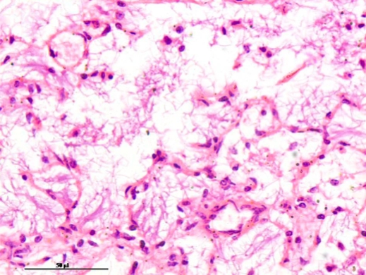
Meconium aspiration syndrome (MAS) is caused due to respiratory distress to the fetus during or before parturition. As a result of asphyxia, the fetal gasping causes meconic amniotic fluid entry into the respiratory air passages and pulmonary complications, and sometimes death of the newborn. In the present study, two cases of MAS in Murrah buffalo calves born full term by assisted delivery and died after few hours of delivery were received for routine postmortem examination. Necropsy examination revealed diffuse mosaic-pattern - like lesions in the lungs characterized by dark brown-to-red deflated (atelectatic) lobes and small irregular pale raised multi focal partially inflated tiny areas amidst the atelectatic lobes. The respiratory passages were filled with aspirated substances. Microscopically, bronchoalveloar lumen contained with keratin, squames and subtle meconium along with mild infiltration of neutrophils in the alveolar parenchyma. The MAS in Murrah buffalo calves in the present study was similar in presentation with that reported in human babies due to the MAS. These cases add further information on MAS in the buffaloes to the scanty literature on this condition.
Downloads
Metrics
References
Castro-Nájera, J.A., J. Martínez-Burnes, D. Mota-Rojas, H. Cuevas-Reyes, A. López, R. Ramírez-Necoechea, R. Gallegos-Sagredo, and M. Alonso-Spilsbury. 2006. Morphological changes in the lungs of meconium-stained piglets. J. Vet. Diagn. Invest., 18(6): 622-627. DOI: 10.1177/104063870601800621
Dargaville, P.A. 2012. Respiratory support in meconium aspiration syndrome: A practical guide. International Journal of Pediatrics, 2012: 1-9. DOI: 10.1155/2012/965159
Davis, R.O., J.B. Philips, B.A. Harris, E.R. Wilson and J.F. Huddleston. 1985. Fatal meconium aspiration syndrome occurring despite airway management considered appropriate. Am. J. Obstet. Gynecol., 151(6): 731-736. DOI: 10.1016/0002-9378(85)90506-x
Dubielzig, R.R. 1977. Pulmonary lesions of neonatal foals. Equine Med. Surg., 1: 419-425.
Goel, A. and S. Nangia. 2017. Meconium aspiration syndrome: challenges and solutions. Research and Reports in Neonatology, 2017(7): 19-28. DOI: 10.2147/RRN.S78106
Lindenskov, P.H., A. Castellheim, O.D. Saugstad and T.E. Mollnes. 2015. Meconium aspiration syndrome: Possible pathophysiological mechanisms and future potential therapies. Neonatology, 107(3): 225-230. DOI: 10.1159/000369373
Lopez, A. and R. Bildfell. 1992. Pulmonary inflammation associated with aspirated meconium and epithelial cells in calves. Vet. Pathol., 29: 104-111. DOI: 10.1177/030098589202900202
Luna, L.G. 1968. Manual of Histologic Staining Methods of the Armed Forces Institute of Pathology, 3rd ed. McGraw-Hill, Toronto, Canada. p. 82-88, 174-188.
McKenzie, H.C. III 2018. Disorders of foals. Equine Internal Medicine, 2018: 1365-1459. DOI: 10.1016/B978-0-323-44329-6.00020-6
Mellor, D.J. and K.J. Stafford. 2004. Animal welfare implications of neonatal mortality and morbidity in farm animals. Vet. J., 168(2):118-133. DOI: 10.1016/j.tvjl.2003.08.004
Newman, S.J., O.O. Fasina. 2019. Meconium aspiration pneumonia and otitis media in two goat kids. J. Vet. Diagn. Invest., 31(3): 463-466. DOI: 10.1177/1040638719834602
Piza, J., M. Gonzalez, C.C. Northrop and R.D. Eavey. 1989. Meconium contamination of the neonatal middle ear. J. Pediatr., 115(6): 910-914. DOI: 10.1016/s0022-3476(89)80741-3
Tanaka, M., T. Izawa, M. Kuwamura, M. Ozaki, T. Nakao, S. Ito and J. Yamate. 2013. A case of meconium aspiration syndrome in a bottlenose dolphin (Tursiops truncatus) calf. J. Vet. Med. Sci., 76(1): 81-84. DOI: 10.1292/jvms.13-0227
Downloads
Published
How to Cite
Issue
Section