Surgical management of fibroadenoma in udder with special reference to ultrasonographic and histopathological findings in three Jaffarabadi buffalo heifers
DOI:
https://doi.org/10.56825/bufbu.2024.4334168Keywords:
Bubalus bubalis, buffaloes, fibroadenoma, Jaffarabadi heifer, mammary gland, ultrasonographyAbstract
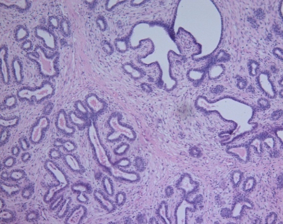
The present report describes a surgical management of fibroadenoma in udder of three Jaffarabadi buffalo heifers of 2-3 years of age. The history of firm udder mass increasing gradually since last 5-6 months. Ultrasonography revealed a mass with isoechoic to hyperechoic parenchyma, separated from surrounding tissue by well differentiated hypoechoic margin. Under sedation and local analgesia, homogenous mass was removed surgically in lateral recumbency. All animals had a postoperative recovery without any complications. Grossly, mass was pale, firm, encapsulated and fasciculated. On histology, tubules and acini lined by layers of cuboidal epithelium and separated by bands of fibrous connective tissue. On the basis of history, clinical findings, ultrasonography and histopathology; these cases were diagnosed as fibroadenoma.
Downloads
Metrics
References
Annapurna, P., V.R. Devi, Md. Riyazuddin and N.S. Babu. 2003. A case of fibroadenoma of mammary gland in a buffalo heifer. Buffalo Bull., 22(4):78-79. Available on: https://kukrdb.lib.ku.ac.th/journal/index.php?/BuffaloBulletin/search_detail/result/286093
Brito, M.F., G.S. Seppa, L.G. Teixeira, T.G. Rocha, T.N. Franca, T.M. Hess and P.V. Peixoto. 2008. Mammary adenocarcinoma in a mare. Cienc. Rural, 38(2): 556-560. DOI: 10.1590/S0103-84782008000200045
Cassali, G.D., G.E. Lavalle, De Nardi, A.B. 2011. Consensus for the diagnosis, prognosis and treatment of canine mammary tumors. Brazilian Journal of Veterinary Pathology, 4(2): 153-180. Available on: https://bjvp.org.br/wp-content/uploads/2015/07/DOWNLOAD-FULL-ARTICLE-29-20881_2011_7_11_14_42.pdf
De Sant’Ana, F.J.F., F.C. Carvalho, C. De O. Gamba, G.D. Cassali, F. Riet-Correa and A.L. Schild. 2014. Mammary diffuse fibroadenomatoid hyperplasia in water buffalo (Bubalus bubalis): Three cases. J. Vet. Diagn. Invest., 26(3):453-456. DOI: 10.1177/1040638714526595
El-Shafaey, E.S. and M. Hamed. 2017. Ultrasonographic and histopathological findings of mammary fibroadenoma in a buffalo heifer (Bubalus bubalis) with special reference to surgical treatment: A case report, Journal of Veterinary Medicine and Allied Science, 1(1): 35-37. Available on https://www.alliedacademies.org/articles/Ultrasonographic%20and%20histopathological%20findings%20of%20mammary%20fibroadenoma%20in%20a%20buffalo%20heifer%20(Bubalus%20bubalis)%20with%20special%20reference%20to%20surgical%20treatment_%20a%20case%20report.pdf
Joshi, D.V., B.J. Patel and S.N. Sharma. 1994. Fibropapilloma of mammary gland in a buffalo calf. Indian Journal of Veterinary Pathology, 18(1): 61-62.
Luna, L.G. 1968. Routine staining procedures: Hematoxylin and Eosin stains. In Manual of Histologic Staining Methods of the Armed Forces Institute of Pathology, 3rd ed. McGraw-Hill, New York, USA.
Mihevc, S.P. and P. Dovc. 2013. Mammary tumors in ruminants. Acta Agriculturae Slovenica, 102(2): 83-86.
Mina, R.B., K. Uchida, A. Sakumi, R. Yamaguchi, S. Tateyama, H. Ogawa and H. Otsuka. 1994. Mammary fibroadenoma in a young Holstein cow. J. Vet. Med. Sci., 56(6): 1171-1172. DOI: 10.1292/jvms.56.1171
Moe, L. 2001. Population-based incidence of mammary tumors in some dog breeds. J. Rep. Fer. S., 57: 439-443.
Raval, S.H., D.V. Joshi, P. Sutariya, B.J. Patel, J.G. Patel and K.R. Joshi. 2015. Fibroadenoma of mammary gland in a Mehsana buffalo. Indian J. Vet. Patho., 39(4): 347-348. DOI: 10.5958/0973-970X.2015.00084.X


.png)


