Cystadenocarcinoma of Bartholin’s gland - Histopathological and immunohistochemical study
DOI:
https://doi.org/10.56825/bufbu.2022.4134265Keywords:
Bubalus bubalis, buffaloes, Bartholin’s gland, Murrah buffalo, cytokeratin, cystadenocarcinomaAbstract
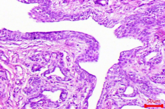
Bartholin’s glands are bilateral, mucus-secreting, tubuloacinar glands located within the submucosa of the vulva of ruminants. Seven years old graded Murrah buffalo was reported to have vaginal prolapse. Clinical examination of the vagina revealed a cyst like structure measuring about 7x6 cm and seen on the Bartholin’s gland and on incision it was pink, soft, and fluctuating. The mass was removed as per the standard routine surgical procedure. The mass had a thick wall and contained about 100 ml of clear fluid. The inner side revealed a rough surface and multiple soft raised areas. Microscopical examination revealed invasive, pleomorphic neoplastic epithelial cells that formed irregular tubules and glands with abundant fibrous stroma and inflammation. The tubules were lined by single to double layered eosinophilic anaplastic cuboidal to columnar cells. The proliferated lining cells had large round to oval vesicular nuclei and prominent nucleoli and a few mitotic figures. Some glandular structures were dilated and cystic or irregularly shaped. Local invasion into the surrounding muscles was noticed. Based on gross and histopathological features the case was diagnosed as Bartholin’s gland cystadenocarcinoma. Immunohistochemical staining revealed strong positive reaction with cytokeratin. After treatment, animal recovered uneventfully and there was no recurrence.
Downloads
Metrics
References
Fathalla, M., M.S. Abdou and H. Fahmi. 1978. Case report: Bartholin gland cyst in the cow. Canadian Vet. J., 19(12): 340. Available on: http://europepmc.org/backend/ptpmcrender.fcgi?accid=PMC1789440&blobtype=pdf
Fathalla, M., N. Hailat, S.Q. Lafi, E.A. Basha and A. Al-Sahli. 2000. An abattoir survey of gross reproductıve abnormalıtıes in the bovine genital tract in Northern Jordan. Isr. J. Vet. Med., 55: 56-61.
Jubb., K. and Palmer’s. 2007. Female Genital System, Pathology of Domestic Animals, 5th ed., Elsevier, Philadelphia, USA.
Manokaran, S., K. Sivasankar, M. Palanisamy, M. Selvaraju and R.E. Napolean, 2014. Unilateral Bartholin’s gland cyst in a Holstein Friesian crossbred cow. International Journal of Livestock Research, 4(9): 48-50. DOI: 10.5455/ijlr.20141120091023
Meuten, D.J. 2017. Tumours of the genital system. Tumours in Domestic Animals, 5th ed. Ames, Iowa, USA. 710p.
Moreira, J.R., T.E.S.D. Oliveira, E.A.D.O. Tongu, R.O. Leite, G.M. Nogueira, M.D.M.Z. Michelazzo, D.J.Z. Delfiol, J.P.E. Saut and S.A. Headley. 2018. Bartholin’s gland adenoma in a Saanen goat. Ciênc. Rural, 48(1). DOI: 10.1590/0103-8478cr20170214
Roberts, J.S. 1971. Veterinary Obstetrics and Genital Diseases, 2nd ed. CBS Publishers and Distributors, New Delhi, India.
Sosnik, H., K. Sosnik and A. Halon. 2007. The pathomorphology of Bartholin’s gland. Analysis of surgical data. Polish Journal Pathology, 58(2): 99-103.
Tanimoto, T., K. Fukunaga and Y. Ohtsuki. 1994. Adenocarcinoma of the major vestibular gland in a cow. Vet. Pathol., 31(2): 246-247. DOI: 10.1177/030098589403100212
Downloads
Published
How to Cite
Issue
Section