Applications of transabdominal ultrasonography in bovine reproduction: A review
DOI:
https://doi.org/10.56825/bufbu.2022.4122365Keywords:
Bubalus bubalis, buffaloes, transabdominal ultrasonography, bovine, reproductionAbstract
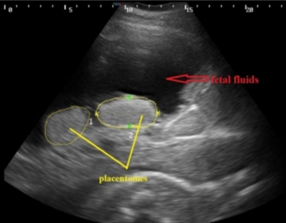
The non-invasive nature of ultrasonography makes it an important clinical and research tool in bovine reproduction and obstetrics. Ultrasound techniques are becoming increasingly important in animal reproduction and obstetrics and these techniques are helpful in evaluation of various disease conditions of uterus during advance pregnancy in bovines. Accordingly, understanding the use of ultrasound technology is of utmost importance in animal sciences, since ultrasound examinations are now a routine part of diagnostic workups in reproduction and obstetrics. Up to now, most of studies were conducted to assess the status of fetal and uterine structures by trans-rectal ultrasonography, but sometimes it was found to be inefficient in judging the complete status of fetal and uterine structures during mid and advance pregnancy when the fetus is located deep into the abdominal cavity. So, in modern times transabdominal ultrasonography is getting popularity over trans-rectal ultrasonography in bovines during mid and advance gestation to determine the complete status of pregnant animals. The new information that has been generated by use of transabdominal ultrasonography has thrown light on diagnostic and research aspects and created new areas for research. Transabdominal ultrasonographic examination of the reproductive tract can provide useful information related to fetal viability, diagnosis of mid to late term pregnancies, gestational age, assessment of fetus and fetal organs, placentomes, fetal fluids, uterine wall, umbilicus, and identification of developmental abnormalities. To best of my knowledge this is the first review of applications of transabdominal ultrasonography in bovine reproduction because literature related to transabdominal ultrasonography in bovine reproduction and obstetrics is very scanty.
Downloads
Metrics
References
Aziz, D.M. 2013. Clinical application of a rapid and practical procedure of transabdominal ultrasonography for determination of pregnancy and fetal viability in cows. Asian Pacific Journal of Reproduction, 2(4): 326-329. DOI: 10.1016/S2305-0500(13)60172-4
Bertolini, M., J.B. Mason, S.W. Beam, G.F. Carneiro, M.L. Sween, D.J. Kominek, A.L. Moyer, T.R. Famula, R.D. Sainz and G.B. Anderson. 2002. Morphology and morphometry of in vivo and in vitro- produced bovine concepti from early pregnancy to term and association with high birth weights. Theriogenology, 58(5): 973-994. DOI: 10.1016/s0093-691x(02)00935-4
Bergamaschi, M.A.C.M., W.R.R. Vicente, R.T. Barbosa, R. Machado, J.A. Marques and A.R. Freitas. 2004. Ultrasound assessment of fetal development in Nelore cows. Arch. Zootec., 53: 371-374. Available on: https://www.redalyc.org/pdf/495/49520404.pdf
Bucca, S. 2006. Diagnosis of the compromised equine pregnancy. Vet. Clin. N. Am., 22(3): 749-761. DOI: 10.1016/j.cveq.2006.07.006
Buczinski, S., G. Fecteau and R.C. Lefebvre. 2007a. Fetal well-being assessment in bovine near-term gestations: current knowledge and future perspectives arising from comparative medicine. Can. Vet. J., 48(2): 178-183.
Buczinski, S., A.M. Belanger, G. Fecteau and J.P. Roy. 2007b. Prolonged gestation in two Holstein cows: Transabdominal ultrasonographic findings in late pregnancy and pathologic findings in the fetuses. J. Vet. Med. A., 54(10): 624-626. DOI: 10.1111/j.1439-0442.2007.00985.x
Buczinski, S. 2009. Ultrasonographic assessment of late term pregnancy in cattle. Vet. Clin. N. Am.-Food A., 25(3): 753-765. DOI: 10.1016/j.cvfa.2009.07.005
Devender., R.K. Chandolia, A.K. Pandey, V. Yadav, P. Kumar and J. Dalal. 2016. Transabdominal color Doppler ultrasonography: A relevant approach for assessment of effects of uterine torsion in buffaloes. Vet. World, 9(8): 842-849. DOI: 10.14202/vetworld.2016.842-849
Devoe, L.D. 2008. Antenatal fetal assessment: contraction stress test, nonstress test, vibroacoustic stimulation, amniotic fluid volume, biophysical profile, and modified biophysical profile-an overview. Semin. Perinatol., 32(4): 247-252. DOI: 10.1053/j.semperi.2008.04.005
England, G.C.W. 1998. Ultrasonographic assessment of abnormal pregnancy. Vet. Clin. N. Am. Small, 28(4): 849-868. DOI: 10.1016/s0195-5616(98)50081-2
Hunnam, J.C., T.J. Parkinson and S. MacDougall. 2009. Transcutaneous ultrasound over the right flank to diagnose mid- to late-pregnancy in the dairy cow. Aust. Vet. J., 87(8): 313-317. DOI: 10.1111/j.1751-0813.2009.00458.x
Hussein, H.A. 2013. Validation of color Doppler ultrasonography for evaluating the uterine blood flow and perfusion during late normal pregnancy and uterine torsion in buffaloes. Theriogenology, 79(7): 1045-1053. DOI: 10.1016/j.theriogenology.2013.01.021
Jonker, F.H., H.A. Van Oord, H.P. Van Geijn, G.C. van der Weijden and M.A. Taverne. 1994. Feasibility of continuous recording of fetal heart rate in the near term bovine fetus by means of transabdominal Doppler. Vet. Quart., 16(3): 165-168. DOI: 10.1080/01652176.1994.9694442
Jonker, F.H. 2004. Fetal death: comparative aspects in large domestic animals. Anim. Reprod. Sci., 82-83(3): 415-430. DOI: 10.1016/j.anireprosci.2004.05.003
Kahn, W. 1989. Sonographic fetometry in the bovine. Theriogenology, 3(5): 1105-1121. DOI: 10.1016/0093-691x(89)90494-9
Kahn, W. 2004. Veterinary Reproductive Ultrasonography. Schlutersche Verlagsgesellschaft MBH and Co., Hannover, Germany. p. 143-168.
Kohan-Ghadr, H.R., R.C. Lefebvre, G. Fecteau, L.C. Smith, B.D. Murphy, J. Sujuki Junior, C. Girard and P. Helie. 2008. Ultrasonographic and histological characterization of the placenta of somatic nuclear transfer-derived pregnancies in dairy cattle. Theriogenology, 69(2): 218-230. DOI: 10.1016/j.theriogenology.2007.09.028
Lawrence, K.E., F.D. Adeyinka, R.A. Laven and G. Jones. 2016. Assessment of the accuracy of estimation of gestational age in cattle from placentome size using inverse regression. New. Zeal. Vet. J., 64(4): 248-252. DOI: 10.1080/00480169.2016.1157050
Lazim, E.H., H.M. Alrawi and D.M. Aziz. 2016. Relationship between gestational age and transabdominal ultrasonographic measurements of fetus and uterus during the 2nd and 3rd trimester of gestation in cows. Asian Pacific Journal of Reproduction, 5(4): 326-330. DOI: 10.1016/j.apjr.2016.06.010
Lerner, J.P. 2004. Fetal growth and well-being. Obstet. Gyn. Clin. N. Am., 31(1): 159-176. DOI: 10.1016/S0889-8545(03)00121-9
Noakes, D.E., T.J. Parkinson and G.C.W. England. 2001. Arthur’s Veterinary Reproduction and Obstetrics, 8th ed. WB Saunders, London, UK.
Reef, V.B. 2007. Late term pregnancy monitoring, p. 410-416. In Samper, J.C., J.F. Pycock and A.O. McKinnon (eds.) Current Therapy in Equine Reproduction. Saunders Elsevier, Saintt Louis, Missouri, USA.
Ribadu, A.Y. and T. Nakao. 1999. Bovine reproductive ultrasonography: A review. J. Reprod. Develop., 45(1): 13-28. DOI: 10.1262/jrd.45.13
Szenci, O., G. Gyulai, P. Nagy, L. Kovacs, J. Varga and M.A. Taverne. 1995. Effect of uterus position relative to the pelvic inlet on the accuracy of early bovine pregnancy diagnosis by means of ultrasonography. Vet. Quart., 17(1): 37-39. DOI: 10.1080/01652176.1995.9694528
Ward, V.L., J.A. Estroff and H.T. Nguyen. 2006. Fetal sheep development on ultrasound and magnetic resonance imaging: A standard for the in-utero assessment of models of congenital abnormalities. Fetal. Diagn. Ther., 21(5): 444-457. DOI: 10.1159/000093888


.png)


