Ultrasonographic characteristics of prostate and seminal vesicle glands in developing Murrah buffalo bulls
DOI:
https://doi.org/10.56825/bufbu.2023.4233507Keywords:
Bubalus bubalis, buffaloes, dimensions, Murrah bull, prostate gland, seminal vesicle, ultrasonographyAbstract
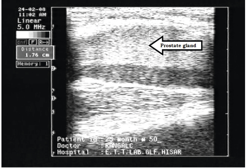
The study was performed in Murrah buffalo-bull calves to assess the developmental dimensions of the prostate and seminal vesicle (SV) glands from 1 to 30 months of age with transrectal ultrasonography. The width of prostate gland ranged between 1.39±0.07 and 2.65±0.14 cm. The rate of change in the mean width of the gland was gradual and almost same from one to thirty month (P<0.05). The correlation co-efficient between width of prostate gland and age was r2 = 0.73. The mean circumference and length of the SV gland at one month to thirty months of age ranged from 6.14±0.32 to 13.05±0.34 cm and 2.61±0.09 to 6.02±0.27 cm, respectively. The correlation co-efficient between length and age, width and age, and circumference of SV gland and age were r2 = 0.68, r2 = 0.55 and r2 = 0.74, respectively. From this study, it is concluded that the ultrasonographic evaluation of prostate and SV along with growth rate could be very practical tool for the assessment of puberty.
Downloads
Metrics
References
Ahmad, N. and D.E. Noakes. 1995. Ultrasound imaging in determining the presence of testicular degeneration in two male goats. Brit. Vet. J., 151(1): 101-110. DOI: 10.1016/s0007-1935(05)80069-7
Ali, M.K., N. Ahmad, N. Akhtar, S. Ali, M. Ahmad and M. Younis. 2011. Ultrasound imaging of testes and epididymides of normal and infertile breeding bulls. Pak. Vet. J., 31(4): 345-350.
Bernardes, O. 2007. Buffaloes breeding in Brazil: Position and economic relevancy. Revista Brasileira de Reprodução Animal, 31: 293-298.
Camela, E.S.C., R.P. Nociti, V.J.C. Santos, B.I. Macente, G.S. Maciel, M.A.R. Feliciano, W.R.R. Vicente, I. Gill, P.M. Bartlewski and M.E.F. Oliveira. 2017. Ultrasonographic characteristics of accessory sex glands and spectral Doppler indices of the internal iliac arteries in peri- and post-pubertal Dorper rams raised in a subtropical climate. Anim. Reprod. Sci., 184: 29-35. DOI: 10.1016/j.anireprosci.2017.06.010
Cartee, R.E., B.W. Grey, T.A. Powe, R.S. Hudson and J. Whitesides. 1989. Preliminary implications of B-mode ultrasonography of the testicles of beef bulls with normal breeding soundness examination. Theriogenology, 31(6): 1149-1157. DOI: 10.1016/0093-691x(89)90083-6
Chandolia, R.K., A. Honaramooz, B.C. Omeke, R. Pierson, A.P. Beard and N.C. Rawlings. 1997. Assessment of development of the testes and accessory glands by ultrasonography in bull calves and associated endocrine changes. Theriogenology, 48(1): 119-132. DOI: 10.1016/S0093-691X(97)00195-7
Chandolia, R.K., G. Singh, A. Kumar, R. Dutt and D.K. Tiwari. 2018. Testicular microlithiasis in a buffalo bull - A rare case. Haryana Veterinarian, 57(1): 122-123. Available on: https://www.luvas.edu.in/haryana-veterinarian/download/harvet2018-june/40.pdf
El-Khawaga, A.R.M., M.M.M. Kandiel, G.A. Sosa, M.E.A.A. El-Roos, A.E. Abdel-Ghaffar and A.E.S. El-Azab. 2012. Ultrasound imaging of the testes and accessory sex glands in buffalo bulls treated with gonadotrophic releasing hormone. J. Reprod. Infertil., 3(1): 8-16. DOI: 10.5829/idosi.jri.2012.3.1.63186
Gnemmi, G. and R.C. Lefebvre. 2009. Ultrasound imaging of the bull reproductive tract: An important field of expertise for veterinarians. Vet. Clin. N. Am. Food A., 25(3): 767-779. DOI: 10.1016/j.cvfa.2009.07.006
Gouletsou, P.G., G.S. Amiridis, P.J. Cripps, T. Lainas, K. Deligiannis, P. Saratsis and G.C. Fthenakis. 2003. Ultrasonographic appearance of clinically healthy testicles and epididymides of rams. Theriogenology, 59(9): 1959-1972. DOI: 10.1016/s0093-691x(02)01259-1
Kastelic, J.P. and L.F.C. Brito. 2012. Ultrasonography for monitoring reproductive function in the bull. Reprod. Domest. Anim., 47(Suppl. 3): 45-51. DOI: 10.1111/j.1439-0531.2012.02042.x
Livestock Census. 2019. 20th Livestock Census, Department of Animal Husbandry and Dairying. Available on: https://dahd.nic.in/sites/default/filess/Key%20Results%2BAnnexure%2018.10.2019.pdf
Manda, S., S. Makkena, B.R. Kakani, S. Chintamaneni and S.N. Kakarla. 2012. Diagnostic applications of ultrasonography to testes and accessory sex glands in Ongole (Bos indicus) bulls. Journal of Advanced Veterinary Research, 2(4): 239-243. Available on: https://www.advetresearch.com/index.php/AVR/article/view/189/186
Pechman, R.D. and B.E. Eilts. 1987. B-mode ultrasonography of the bull testicle. Theriogenology, 27(2): 431-443. DOI: 10.1016/0093-691x(87)90231-7
Ranga, L.C., R.K. Chandolia, S.K. Phulia and L. Singh. 2014. Ultrasono graphic measurements of bulbo urethral glands in developing Murrah buffalo. Indian Journal of Animal Reproduction, 35(1): 14-17.
Ribadu, A.Y. and T. Nakao. 1999. Bovine reproductive ultrasonography: A review. J. Reprod. Develop., 45(1): 13-28. Available on: https://www.jstage.jst.go.jp/article/jrd/45/1/45_1_13/_pdf/-char/en
Rodrigues, N.N., G.F. Rossia, D.P. Vrismana, A.R. Tairaa, L.L. Souzab, M.F. Zorzettob, N.M. Bastosa, C.C.P. de Pazb, V.F.M.H. de Limaa, F.M. Monteirob and M.E.F. Oliveiraa. 2020. Ultrasonographic characteristics of the testes, epididymis and accessory sex glands and arterial spectral indices in peri- and postpubertal Nelore and Caracu bulls. Anim. Reprod. Sci., 212: 1-10. DOI: 10.1016/j.anireprosci.2019.106235
Schnobrich, M.R., R.O. Turner, C.N. Belcher and J. Slack. 2015. Transrectal ultrasonographic characterization of the accessory sex glands, pelvic urethra, and ureters in normal geldings. Theriogenology, 85(2): 186-192. DOI: 10.1016/j.theriogenology.2015.09.008
Singh, K., A. Kumar, M. Honparkhe and D. Dadarwal. 2015. Ultrasonographic approaches for breeding soundness evaluation of high and low libido buffalo bulls. Indian J. Anim. Sci., 85(5): 451-453.
Sunder, S., U. Singh, R.K. Chandolia and J. George. 2013. Diagnosis of prostate hyperplasia in a dog using 3D/4D ultrasonography. Haryana Vet., 52: 133-134. Available on: https://www.luvas.edu.in/haryana-veterinarian/download/harvet2013/39.pdf
Weber, J.A., C.J. Hilts and G.L. Woods. 1988. Ultrasonographic appearance of bull accessory sex glands. Theriogenology, 29(6): 1347-1353. DOI: 10.1016/0093-691X(88)90015-5
Downloads
Published
How to Cite
Issue
Section