Ultrasonography and biochemical studies of hepatobiliary system in buffaloes
DOI:
https://doi.org/10.56825/bufbu.2022.4123957Keywords:
Bubalus bubalis, buffaloes, hepatobiliary, biochemical studies, ultrasonographyAbstract
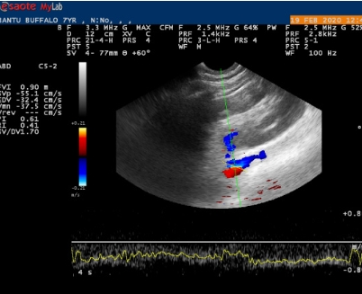
The present work was undertaken to study the ultrasonographic and clinico-biochemical parameters of hepatobiliary system in apparently healthy adult non gravid buffaloes. The present study revealed that, the hepatic parenchyma was homogenously coarse echogenic, interspersed with anechoic bands of hepatic vessels, with sharp margin and was hyperechoic relative to right renal cortex. It was imaged from just behind the last rib to the 6th intercostal space. The gallbladder was visualized between the 12th to 9th intercostal spaces, seen as a pear-shaped fluid-filled anechoic structure with hyperechoic wall, restricted to one to two intercostal spaces. The portal vein was seen as a stellate, branching, anechoic structure with hyperechoic wall and the hepatic vein as anechoic tubular structure with anechoic wall within the hepatic parenchyma. The caudal vena cava was observed as a triangular anechoic structure in transverse view and tubular in longitudinal view. It is concluded that the ultrasonography is a useful tool for the non-invasive examination of liver. Its sonographic appearance and parameters measured in healthy buffaloes can serve as reference values for the diagnosis of pathological changes in liver.
Downloads
Metrics
References
Abu-Yousef, M.M. 1992. Normal and respiratory variations of the hepatic and portal venous duplex doppler waveforms with simultaneous electrocardiographic correlation. Journal of Medical Ultrasound, 11(6): 263-268. DOI: 10.7863/jum.1992.11.6.263
Acorda, J.A., M.H. Yamada and S.M. Ghamsari. 1994. Evaluation of fatty infiltration of the liver in dairy cattle through digital analysis of hepatic ultrasonograms. Veterinary Radiology and Ultrasound, 35(2): 120-123. DOI: 10.1111/j.1740-8261.1994.tb00199.x
Ahrens, F.A. 1967. Histamine, lactic acid, and hypertonicity as factors in the development of rumenitis in cattle. Am. J. Vet. Res., 28(126): 1335-1342.
Belotta, A.F., B.P. Santarosa, D.O.L. Ferreira, S.M.F. Carvalho, R.C. Gonçalves, C.R. Padovani and M.J. Mamprim. 2017. Portal Vein Doppler flow metry in healthy sheep according to age. Brazilian Journal of Veterinary Research and Animal Science, 37(10): 1172-1176. DOI: 10.1590/s0100-736x2017001000021
Braun, U. 1990. Ultrasonographic examination of the liver in cows. Am. J. Vet. Res., 51(10): 1522-1526.
Braun, U. 2009a. Ultrasonography of the gastrointestinal tract in cattle. Vet. Clin. N. Am-Food A., 25(3): 567-590. DOI: 10.1016/j.cvfa.2009.07.004
Braun, U. 2009b. Ultrasonography of the liver in cattle. Vet. Clin. N. Am.-Food A., 25(3): 591-609. DOI: 10.1016/j.cvfa.2009.07.003
Braun, U. and D. Gerber. 1994. Influence of age, breed, and stage of pregnancy on hepatic ultrasonographic findings in cows. Am. J. Vet. Res., 55(9): 1201-1205.
Dirksen, G. 1979. Clinical Examination of Cattle. Verlag Paul Parey, Berlin, Germany.
Imran, S. 2010. Ultrasonography of bovine abdominal cavity. M.V.Sc. Thesis, Chaudhary Sarwan Kumar Himachal Pradesh Krishi Vishvavidyalaya Palampur (H.P.), India.
Jackson, P. and P. Cockcroft. 2008. Cattle clinical examination by body system and region, p. 7-216. In Clinical Examination of Farm Animals, 1st ed. John Wiley and Sons, New Delhi, India.
Khalphallah, A., M. Abdelhakiem and E. Elmeligy. 2016. The ultrasonographic findings of the liver, gall bladder and their related vasculatures in healthy Egyptian buffaloes (Bubalus bubalis). Assiut Veterinary Medical Journal, 62(149): 156-162. DOI: 10.21608/AVMJ.2016.169978
Kremer, H., W. Dobrinski and M.A. Schreiber. 1994. In Kremer, H., W. Dobrinski, I. Medizin and A. Gebiete (eds.) Sonographic Diagnostic, 4th ed. Urban and Schwarzenberg, Munich, Germany. 63p.
Kumar, A., N.S. Saini, J. Mohindroo and N.K. Sood. 2012. Ultrasonographic features of normal heart and liver in relation to diagnose pericarditis in bovine. Indian J. Anim. Sci., 82(12): 1489-1494.
Kumar, A., N.S. Saini, J. Mohindroo, B.B. Singh, V. Sangwan and N.K. Sood. 2016. Comparison of radiography and ultrasonography in the detection of lung and liver cysts in cattle and buffaloes. Vet. World, 9(10): 1113-1120. DOI: 10.14202/vetworld.2016.1113-1120
Nyland, T.G., D.A. Hager and D.S. Herring. 1989. Sonography of the liver, gallbladder, and spleen. Semin. Vet. Med. Surg. Small Anim., 4(1): 13-31.
Patel, M.D., A. Lateef, H. Das, M.V. Prajapati, P. Kakati and H.R. Savani. 2016. Estimation of blood biochemical parameters of Banni buffalo (Bubalus bubalis) at different age, sex and physiological stages. J. Livest. Sci., 7: 250-255. Available on: http://livestockscience.in/wp-content/uploads/Biochem-prof-Banni-Buff.pdf
Radostits, O.M., C.C. Gay, K.W. Hinchcliff and P.D. Constable. 2007. A Textbook of the Diseases of Cattle, Sheep, Pigs, Goats and Horses, 10th ed. Saunders, USA.
Rantanen, N.W. 1986. Diseases of the liver. Vet. Clin. N. Am.-Equine, 2: 105-114.
Sangwan, V. 2015. B-mode and doppler sonographic study of major blood vessels in cattle and buffalo. Ph.D. Thesis, Guru Angad Dev Veterinary and Animal Sciences University, Ludhiana (Punjab), India.
Sood, P., A.S. Nanda, N. Singh, M. Javed and R. Prasad. 2009. Effect of lameness on follicular dynamics in crossbred cows. Veterinary Archives, 79(2): 131-136. Available on: https://hrcak.srce.hr/file/58670
Starke, A., S. Schmidt, A. Haudum, T. Scholbach, P. Wohlsein, M. Beyerbach and J. Rehage. 2011. Evaluation of portal blood flow using transcutaneous and intraoperative Doppler ultrasonography in dairy cows with fatty liver. J. Dairy Sci., 94(6): 2964-2971. DOI: 10.3168/jds.2011-4156
Streeter, R.N. and D.L. Step. 2007. Diagnostic ultrasonography in ruminants. Vet. Clin. N. Am.-Food A., 23(3): 541-574. DOI: 10.1016/j.cvfa.2007.07.008
Verma, Y. and M. Swamy. 2009. Prevalence and pathology of hydatidosis in buffalo liver. Buffalo Bull., 28(4): 207-211.


.png)


