
กุญแจสู่ความสำเร็จในการตรวจตาในสุนัขและแมว | Keys to a successful canine and feline ocular exam
Article Sidebar
Main Article Content
Abstract
บทคัดย่อ
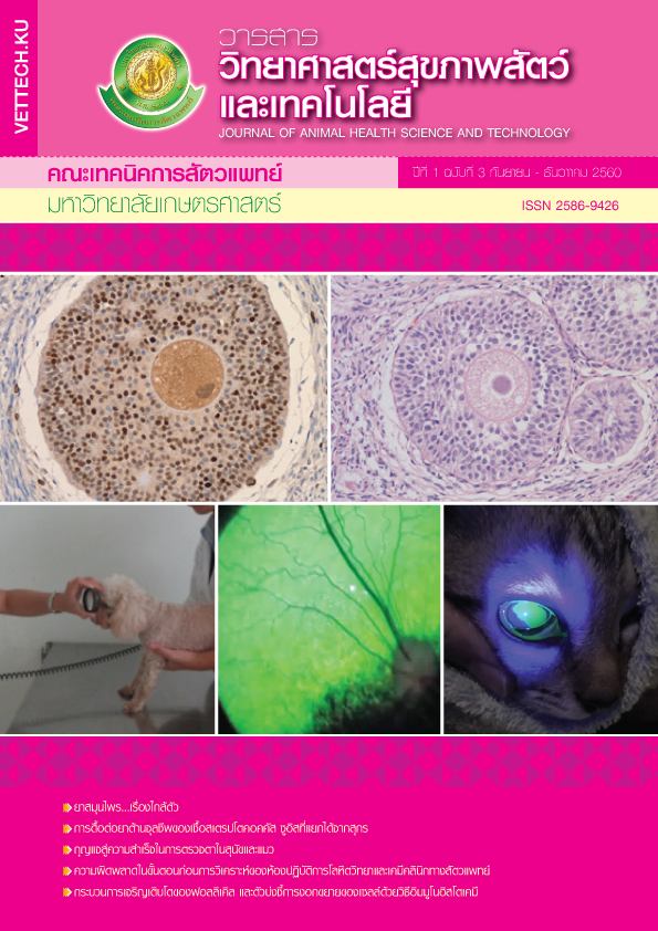
ก่อนที่สัตว์ป่วยจะได้รับการรักษาโรคทางตาที่เหมาะสม สัตวแพทย์หรือผู้ตรวจจะต้องวินิจฉัยความผิดปกติต่างๆ ที่พบได้อย่างถูกต้อง โดยเมื่อทำการตรวจตาในสุนัขและแมวจะต้องตรวจให้ครบทุกส่วน ได้แก่ หนังตา กระจกตา ห้องหน้าม่านตา แก้วตา เนื้อวุ้นตา และส่วนจอประสาทตา ด้วยเครื่องมือตรวจวินิจฉัยที่สำคัญอันเป็นพื้นฐาน ได้แก่ กระดาษวัดการสร้างน้ำตา เครื่องวัดความดันตา สีย้อมกระจกตา และกล้องส่องตรวจจอตา ซึ่งเครื่องมือช่วยตรวจวินิจฉัยเหล่านี้จำเป็นจะต้องนำมาใช้ในสัตว์ป่วยทุกครั้งที่มีการตรวจตา นอกจากนี้ ลำดับในการตรวจตาก็สำคัญ เพราะการตรวจหรือการสังเกตบางอย่างจะต้องทำก่อนการทดสอบอื่นๆ ขอบเขตของบทความนี้จึงเป็นการบรรยายภาพรวมของขั้นตอนการตรวจตาพื้นฐานที่จะเป็นกุญแจสู่ความสำเร็จในการวินิจฉัยโรคทางตาและช่วยให้ผู้ทำการตรวจมีความสะดวกและมั่นใจในตรวจตาทุกขั้นตอน
Abstract
Before the patient can be properly treated for an ophthalmic condition, the veterinarian or examiner must correctly diagnosis any abnormal ophthalmic finding. When performing an ocular exam in dogs and cats, the examiner must evaluate all areas of eyelid, cornea, anterior chamber, lens, vitreous body and retina. The importance of thorough diagnostic testing such as, Schirmer tear test, intraocular pressure measurement, fluorescein staining, direct and indirect ophthalmoscopy should be performed in all ophthalmic patients. In additions, the order of testing is important because some tests or observations should be performed before others. The scope of this article is to give an overview of the basic steps procedures involved which are the keys to a successful ophthalmic exam and to aid the examiner feel comfortable and confident with the overall process.