COMPARATIVE EVALUATION OF RETICULAR MORPHOMETRY BY ULTRASONOGRAPHY IN HEALTHY WITH RUMINAL INDIGESTION AND FOREIGN BODY SYNDROME AFFECTED BUFFALOES
DOI:
https://doi.org/10.56825/bufbu.2025.4414256Keywords:
Bubalus bubalis, buffaloes, ultrasonography, reticulum, ruminal indigestion, foreign body syndromeAbstract
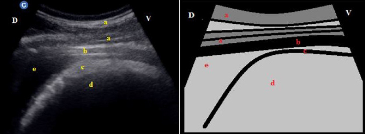
The ultrasonographic study was conducted to evaluate morphometry of reticulum in 32 clinical cases of buffaloes presented to Veterinary College, Bidar. They were divided into three groups, Group 1 (13) healthy buffaloes, Group 2 (13) buffaloes with ruminal indigestion and Group 3 (6) buffaloes with foreign body syndrome. Reticulum sonography was carried out without sedation in standing position at 6th to 8th intercostal space. To know the appearance, contour, motility, wall thickness and distance from abdominal wall to reticulum the four windows were approached. The reticulum is visible at frequency of 3.5 to 4.5 MHz, depth of 14 to 16 cm and gain of 130 to 160 in real time B-mode scanner. In Group 2 buffaloes the appearance of reticulum wall was half moon or crescent shape with smooth contour and significantly decreased reticular motility to 1.30±0.25/4 minutes. In Group 3, the appearance of the right lateral reticulum wall revealed irregular shape with uneven contour in 2 buffaloes. The right lateral reticular wall thickness was significantly increased to 0.95±0.13 cm. Reticular motility was significantly decreased to 1.17±0.15/4 minutes. Distance between abdominal wall to reticulum on right lateral examination was 7.09±1.24 cm. Among the forestomach reticulum is the motile organ so better diagnosed by ultrasonography than other imaging modalities. Pathological signs confirmed by ultrasonography scanning are presence of peritoneal fluid, movement of fibrin shreds, adhesions, abscess and hernia of reticulum in buffaloes affected with foreign body syndrome. In ruminal indigestion, nil to reduced reticular motility is the only important sonographic finding.
Downloads
Metrics
References
Abdelaal, A.M., M. Floeck, S. El Maghawry and W. Baumgartner. 2009. Clinical and ultrasonographic differences between cattle and buffaloes with various sequelae of traumatic reticuloperitonitis. Vet Med., 54(9): 399-406. DOI: 10.17221/128/2009-VETMED
Abdelaal, A.M., M.B. Mostafa, A.M. Abu-Seida, O.S. Al-Abbadi and S.F. Abbas. 2016. Ultrasonographic findings in hardware diseased buffaloes (Babulus babilus). Research Journal of Pharmaceutical Biological and Chemical Sciences, 7(5): 1644-1649. Available on: https://rjpbcs.com/pdf/2016_7(5)/[210].pdf
Abouelnasr, K.S., E. Mosbah, G.I. Karrouf and A.E. Zaghloul. 2012. Comparative ultrasonographic findings of traumatic reticulitis, perireticular abscess and diaphragmatic hernia in buffalo (Bubalus bubalis). Journal of American Science, 8(8): 590-595. Available on: https://www.jofamericanscience.org/journals/am-sci/am0808/090_8843am0808_590_595.pdf
Abouelnasr, K.S., E.M. Mosbah, G.I. Karrouf and A.E. Zaghloul. 2014. Ultrasonography of normal reticulum in 30 healthy buffalo (Bubalus bubalis). J. Appl. Anim. Res., 42(2): 153-159. DOI: 10.1080/09712119.2013.823435
Abu-Seida, A.M. and O.S. Al-Abbadi. 2016. Recent advances in the management of foreign body syndrome in cattle and buffaloes: A review. Pak. Vet. J., 36(4): 385-393.
Athar, H., J. Mohindroo, K. Singh and T. Singh. 2010. Clinical, haematobiochemical, radiographic and ultrasonographic findings in bovines with rumen impaction. Intas Polivet, 11(2): 180-183.
Braun, U. and M. Gotz. 1994. Ultrasonography of the reticulum in cows. Am. J. Vet. Res., 55(3): 325-332.
Braun, U. 2003. Ultrasonography in gastrointestinal disease in cattle. Vet. J., 166(2): 112-124. DOI: 10.1016/s1090-0233(02)00301-5
Khalphallah, A., H.K. Elsayed, E.N.A.S. Elmeligy and S.F. El-Hawari. 2016a. The ultrasonographic findings of the gastrointestinal tract and spleen in healthy Egyptian buffaloes (Bubalus bubalis). Assiut Veterinary Medical Journal, 62(148): 9-47. Available on: https://avmj.journals.ekb.eg/article_169208_e88f5d15dc741d4a308947f1c6a610c5.pdf
Khalphallah, A., A.M. Abu-Seida, M. Abdelhakiem, E. Elmeligy and U.T. Mahmoud. 2016b. Laboratory, radiographic and ultrasonographic findings of acute traumatic reticuloperitonitis in buffaloes (Bubalus bubalis). Asian J. Anim. Vet. Adv., 11(11): 675-683. DOI: 10.3923/ajava.2016.675.683
Khalphallah, A., E. Elmeligy, H.K. Elsayed, S.F. El-Hawari and M.H. Elrashidy. 2016c. Diagnostic significance of ultrasonography in complicated traumatic reticuloperitonitis in Egyptian buffaloes (Bubalus bubalis). Asian J. Anim. Vet. Adv., 11: 319-330. DOI: 10.3923/ajava.2016.319.330
Makhdoomi, S.M., V. Sangwan, A. Kumar, J. Mohindroo and A. Gupta. 2019. Ultrasonographic morphometry of reticulum in cattle and buffaloes suffering from traumatic reticulo-peritonitis. Buffalo Bull., 38(3): 421-436. Available on: https://kukrdb.lib.ku.ac.th/journal/BuffaloBulletin/search_detail/result/390583
Mostafa, M.B., A.M. Abu-Seida, A.M. Abdelaal, O.S. Al-Abbadi and S.F. Abbas. 2015. Ultrasonographic features of the reticulum in normal and hardware diseased buffaloes. Research Opinions in Animal and Veterinary Sciences, 5(4): 165-171.
Sheikh, I., S. Shivali and A.A. Bhat. 2012. Ultrasonographic imaging of normal reticulum and traumatic reticuloperitonitis in crossbred cows. Eurasian J. Vet. Sci., 28: 214-219. Available on: https://scispace.com/pdf/ultrasonographic-imaging-of-normal-reticulum-and-traumatic-38x0e5kb6l.pdf
Snedecor, G.W. and W.G. Cochran. 1994. Statistical Methods, 8th ed. Affiliated East-West Press Pvt Ltd, New Delhi, India. p. 53-58.
Downloads
Published
How to Cite
Issue
Section