Gross as well as microscopic anatomy and physiological functions of fetal placenta in Jaffrabadi buffaloes
DOI:
https://doi.org/10.56825/bufbu.2024.4314022Keywords:
Bubalus bubalis, buffaloes, gross-morphology, histo-morphometry, trophobalst, hormones, placenta, cotyledon, Jaffrabadi buffaloAbstract
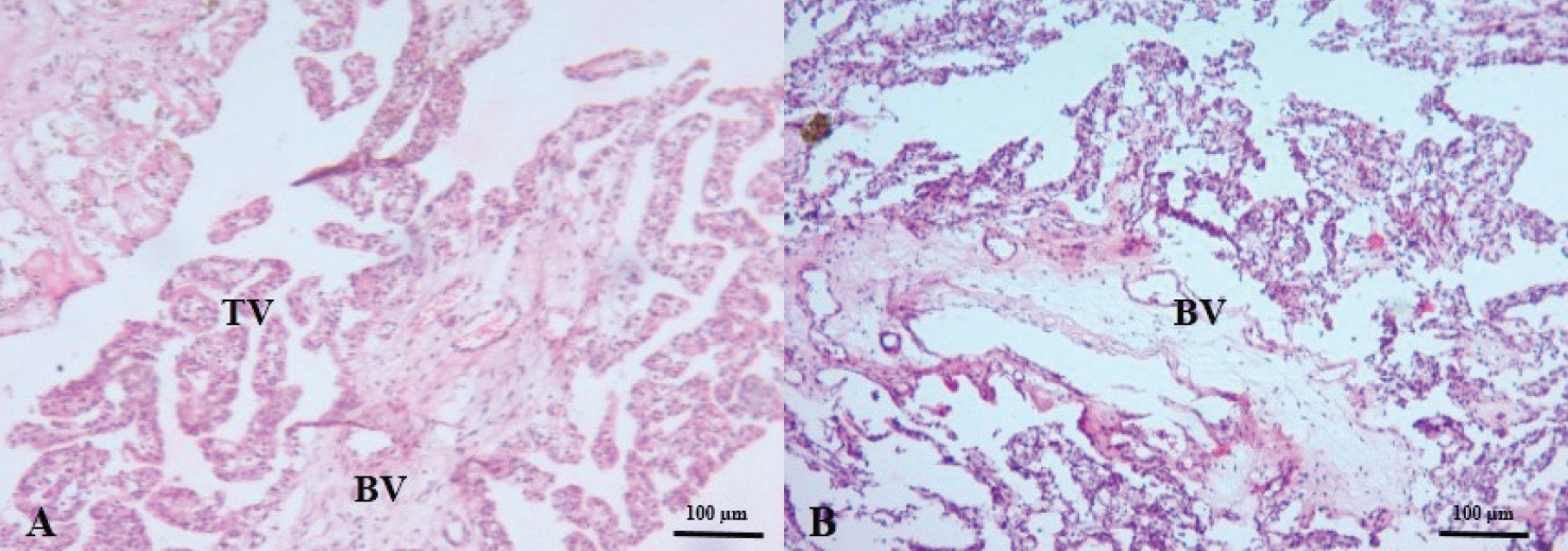
The present study was conducted to know the gross morphology and histo-morphological structure of fetal placenta in Jaffrabadi buffaloes. Parameters like calf weight, placental weight, numbers, and size of cotyledons were observed in Jaffrabadi buffaloes and morphologically, fetal cotyledons were convex and non-pendunculated in Jaffrabadi buffaloes were found. Histological studies of small and large cotyledons showed extensive branching of secondary and tertiary villi that were longer, slender, and well developed in Jaffrabadi buffalo. A less developed basal lamina was seen in small cotyledon whereas developed basal lamina with numerous capillaries and connective tissue were observed in the large cotyledon. The diameter of trophoblast giant cells (TGC) in larger cotyledons were significantly (P<0.05) than the small cotyledons in expelled placenta at full term in Jaffrabadi buffaloes. A distinct distribution of carbohydrate and lipids in cotyledons were observed between large and small cotyledons as evident by acid mucopolysaccharides, neutral polysaccharides, and sudanophilic staining. Specific staining for calcium with Alizarin red stain showed that calcium is not present in a noticeable amount in small and large cotyledons. Isolation and culture of Jaffrabadi placental cells in M-199 medium with antibiotics and 2% FBS results in the efficient production of progesterone, estrogen, and testosterone. This study has shown that trophoblast cells are the actual sites for steroid hormone production. These cultured placental cells (1x10⁶ cells/ ml) produce Progesterone, Estradiol-17β and Testosterone in the range of 1.72 to 2.12, 16.03 to 19.51 and 0.51 to 0.58 ng/ml, respectively in Jaffrabadi buffalo.
Downloads
Metrics
References
Adeyinka, F.D. 2012. The development of the bovine placentome and associated structures during gestation. Ph.D. Thesis, Massey University, Palmerston North, New Zealand.
Agababov, R.M., T.N. Abashina, N.E. Suzina, M.B. Vainshtein and P.M. Schwartsburd. 2007. Link between the early calcium deposition in placenta and nanobacterial-like infection. J. Biosciences, 32(6): 1163-1168. DOI: 10.1007/s12038-007-0118-9
Al-Ramadan, S.Y. 2014. Camel endometrium: Light microscopic and ultrastructural features related to pregnancy. Ruminant Science, 3(2): 129-140.
Andresen, A. 1927. Die Plazentome der Wiederkäuer. Morphologisches Jahrbuch Leipzig, 57: 410-485.
Baur, R. 1972. Quantitative Analyse des Wachstums der Zottenoberfläche bei der Placenta des Rindes und des Menschen. Z. Anat. Entwicklungs., 136(1): 87-97.
Bhuyan, D., J. Dutta, C. Das, S. Sinha, N.K. Sarma and A. Das. 2016. Morphological characteristics of foetal membranes of swamp buffaloes of Assam. Indian J. Anim. Res., 50(2): 156-159. Available on: https://www.cabidigitallibrary.org/doi/pdf/10.5555/20173082763
Bjorkman, N. 1969. Light and electron microscopic studies on cellular alterations in the normal bovine placentome. Anat. Rec., 163(1): 17-29. DOI: 10.1002/ar.1091630103
Bjorkman, N. 1954. Morphological and histochemical studies on the bovine placenta. Acta Anat., 22: 1-91.
Carter, A.M. 2019. Evolution of placentation in cattle and antelopes. Anim. Reprod., 16(1): 3-17. DOI: 10.21451/1984-3143-AR2018-00145
Carvalho, A.F., K. Klisch, M.A. Miglino, F.T.V. Pereira and E. Bevilacqua. 2006. Binucleate trophoblast giant cells in the water buffalo (Bubalus bubalis) placenta. Journal of Morphology, 267(1): 50-56. DOI: 10.1002/jmor.10387
Chayen, J., L. Bitenski, R. Buther and L. Poulter. 1969. Histochemistry: A Guide to Practical Histochemistry, Oliver and Boyd, Edinburg, England.
Duello, T.M., J.C. Byatt and R.D. Bremel. 1986. Immunohistochemical localization of placental lactogen in binucleate cells of bovine placentome. Endocrinology, 119(3): 1351-1355. DOI: 10.1210/endo-119-3-1351
Hegde, N.G. 2019. Buffalo husbandry for sustainable development of small farmers in India and other developing countries. Asian Journal of Research in Animal and Veterinary Sciences, 3(1): 1-20.
Hong, S.H., S.C. Kim, M.N. Park, J.S. Jeong, S.Y. Yang, Y.J. Lee, O.N. Bae, H.S. Yang, S. Seo, K.S. Lee and B.S. An. 2019. Expression of steroidogenic enzymes in human placenta according to the gestational age. Mol. Med. Rep., 19(5): 3903-3911. DOI: 10.3892/mmr.2019.10048
Hradecky, P., H.W. Mossman and G.G. Stott. 1988. Comparative histology of antelope placentomes. Theriogenology, 29(3): 693-729. DOI: 10.1016/S0093-691X(88)80015-3
Igwebuike, U.M. 2006. Trophoblast cells of ruminant placentas-A minireview. Anim. Reprod. Sci., 93(3-4): 185-198. DOI: 10.1016/j.anireprosci.2005.06.003
Khatri, P. 2011. The Bovine placenta as a source and target of steroid hormones: Aspects on the role of Androgens and sulfonated steroids. Ph.D. Thesis, Faculties of Veterinary Medicine and Medicine of the Justus Liebig University, Giessen, Germany.
Kouamo, J., A.M.N. Saague and A.P. Zoli. 2018. Determination of age and weight of bovine fetus (Bos indicus) by biometry. Journal of Livestock Science, 9: 9-15. Available on: https://livestockscience.in/wp-content/uploads/bovinefetalagebiometry.pdf
Kumar, A. and S. Kaur. 2017. Calcium: a nutrient in pregnancy. Journal of Obstetrics and Gynecology of India, 67(5): 313-318. DOI: 10.1007/s13224-017-1007-2
Kumar, R. and R. Singh. 2010. Buffalo production system in India. Revista Veterinaria, 21(Suppl. 1): 32-37.
Lee, T.Y., A.M. Jamieson and I.A. Schafer. 1973. Changes in the composition and structure of glycosaminoglycans in the human placenta during development. Pediatr. Res., 7(12): 965-977. DOI: 10.1203/00006450-197312000-00005
Luna, L.G. 1968. Manual of Histologic Staining Methods of the Armed Forces Institute of Pathology, 3rd ed. McGraw Hill Book Co., New York, USA.
Manzoor, A., H. Khan, I. Maqbool, Z. Dar, T. Akram and M. Banday. 2018. Disorders compromising reproductive health and their therapeutics in dairy animals - A review. International Journal of Livestock Research, 8(3): 24-38. DOI: 10.5455/ijlr.20170501110504
Mossman, H.W. 1987. Vertebrate Foetal Membranes, Rutgers University Press, New Brunswick, Canada.
Murugeppa, A., M.M. Appannavar, M.M. Patil and S.S. Honnappagol. 1998. Study on placental membrane and its effect on subsequent fertility in Surti buffaloes. Buffalo Bull., 17(2): 35-36. Available on: https://kukrdb.lib.ku.ac.th/journal/BuffaloBulletin/search_detail/result/285997
Oliveira, C.M. de, A.B. de Oliveira, E.M. Ramos, T.V. Cavalcante and V.M. Maruo. 2010. Histological characterization of the placenta of zebu cattle raised in Oriental Amazonia, Brazil. Acta Veterinaria Brasilica, 4(2): 100-104.
Padodara, R.J. and J.S. Arya. 2014. Reproductive performance of Triple cross cattle. Journal of Indian Veterinary Association, Kerala, 12(2): 50-52.
Peter, A.T. 2013. Bovine placenta: A review on morphology, components, and defects from terminology and clinical perspectives. Theriogenology, 80(7): 693-705. DOI: 10.1016/j.theriogenology.2013.06.004
Polei, M., J. Günther, D. Koczan and R. Fürbass. 2020. Trophoblast cell differentiation in the bovine placenta: differentially expressed genes between uninucleate trophoblast cells and trophoblast giant cells are involved in the composition and remodeling of the extracellular matrix and O-glycan biosynthesis. BMC Molecular and Cell Biology, 21(1): 1-12. DOI: 10.1186/s12860-020-0246-8
Prasanth Babu, A. 2008. Histological and Histochemical studies on the placentomes of the buffalo (Bubalus bubalis). M.V.Sc. Thesis, Sri Venkateswara Veterinary University, Tirupati, India.
Prophet, E.B., B. Mills, J.B. Arrington and L.H. Sobin. 1994. Laboratory Methods in Histotechnology, American Registry of Pathology, Armed Forces Institute of Pathology, Washington DC, USA. 198p.
Raja. R. 1982. Gross, histological and histochemical studies of placenta in buffalo (Bubalus bubalis). Ph.D. Thesis, C.S.A. University of Agriculture and Technology, Mathura campus, Matura, India.
Ranjan, R. and O. Singh. 2019. Histo-architectural changes in placental epithelium during gestation in buffaloes (Bubalus bubalis). Buffalo Bull., 38(2): 193-201. Available on: https://kukrdb.lib.ku.ac.th/journal/BuffaloBulletin/search_detail/result/388956
Ranjan, R. and O. Singh. 2013. Gross-morphological studies on placentomes of buffalo (Bubalus bubalis). Indian Vet. J., 90(8): 28-31.
Ranjan, R., O. Singh and R.S. Sethi. 2013. Histochemistry of placenta of buffalo (Bubalus bubalis). Indian Vet. J., 90(4): 16-18.
Rodriguez, T.A., D.B. Sparrow, A.N. Scott, S.L. Withington, J.I. Preis, J. Michalicek, M. Clements, T.E. Tsang, T. Shioda, R.S. Beddington and S.L. Dunwoodie. 2004. Cited1 is required in trophoblasts for placental development and for embryo growth and survival. Mol. Cell. Biol., 24(1): 228-244. DOI: 10.1128/MCB.24.1.228-244.2004
Sarvaiya, N.P., V.M. Mehta, V. Deshpande and K. Jankiraman. 1990. Effect of sex of calf, season of calving and parity on placental weight and expulsion time in Surti buffaloes. Cheiron, 19(3): 122-126.
Schmidt, S. 2005. Morphology of peri-partal placentomes and post-partal foetal membranes in African buffalo (Syncerus caffer) and comparative aspects with cattle (Bos taurus). M.Sc. Thesis, University of Pretoria, Pretoria, South Africa.
Schneider, C.A., W.S. Rasband and K.W. Eliceiri. 2012. NIH Image to ImageJ: 25 years of image analysis. Nat. Methods, 9(7): 671-675.
Snedecor, G.W. and W.G. Cochran. 1994. Statistical Methods, 8th ed. Oxford and IBH Publishing Co. Calcutta, India.
Wallingford, M.C., C. Benson, N.W. Chavkin, M.T. Chin and M.G. Frasch. 2018. Placental vascular calcification and cardiovascular health: It is time to determine how much of maternal and offspring health is written in stone. Front. Physiol., 9: 1044. DOI: 10.3389/fphys.2018.01044
Wango, E.O., F.B. Wooding and R.B. Heap. 1990. The role of trophoblastic binucleate cells in implantation in the goat: A morphological study. J. Anat., 171: 241-257.
Wooding, F.B.P. 1992. Current topic: The synepitheliochorial placenta of ruminants: binucleate cell fusions and hormone production. Placenta, 13(2): 101-113. DOI: 10.1016/0143-4004(92)90025-O
Downloads
Published
How to Cite
Issue
Section