Microscopic exploration into the behavior of giant cells in placentomes of buffalo (Bubalus bubalis)
DOI:
https://doi.org/10.56825/bufbu.2023.4235464Keywords:
Bubalus bubalis, buffaloes, placentomes, giant cells, histology, histochemistryAbstract
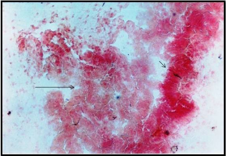
The present study was conducted on placentomes of 31 pregnant buffaloes ranging from 38 to 243 days of gestation to explore the microscopic details of giant cells and their behavior in buffalo placentomes. Various histological and histochemical stains used in the study revealed its structural and chemical details. The study revealed that giant cells played a major role in transplacental transfer of nutrients and other metabolites required by the fetal and maternal tissues. High polysaccharide content and intense enzymatic reaction indicated high metabolic activity of the giant cells in the placenta. The migratory nature of giant cells observed in the present study revealed its role in transfer of metabolites. The cytoplasmic processes observed in the study indicated its fusion with the cryptal epithelium as a medium of transfer of metabolites and formation of multinucleated giant cells. Strong acid phosphates activity can be correlated with its erythrophagocytic nature as a medium of transfer of iron molecules to the developing fetus.
Downloads
Metrics
References
Chayen, J., R.G. Butcher, L. Bitensky and L.W. Poulter. 1969. A Guide of Practical Histochemistry. Oliver and Boyol, Edinbury, England. p. 83-174.
Christie, G.A. 1968. Comparative histochemical studies on carbohydrate, lipid and RNA metabolism in the placenta and foetal membranes. J. Anat., 103(1): 91-112.
Hradecky, P., H.W. Mossman and G.G. Stott. 1987. Comparative development of ruminant placentomes. Theriogenology, 29(3): 715-729. DOI: 10.1016/s0093-691x(88)80016-5
Igwebuike, U.M. 2004. Trophoblast cells of ruminant placentas-A minireview. Anim. Reprod. Sci., 93(3-4): 185-198. DOI: 10.1016/j.anireprosci.2005.06.003
Klisch, K. and L. Leiser. 2003. In bovine binucleate trophoblast giant cells, pregnancy-associated glycoproteins and placental prolactin-related protein-I are conjugated to asparagine-linked N-acetylgalactosaminyl glycans. Histochem. Cell Biol., 119(3): 211-217. DOI: 10.1007/s00418-003-0507-6
Klisch, K., W. Hecht, C. Pfarrer, G. Schuler, B. Hoffmann and R. Leiser. 1999. DNA content and ploidy level of bovine placentomal trophoblast giant cells. Placenta, 20(5-6): 451-458. DOI: 10.1053/plac.1999.0402
Lawn, A.M., A.D. Chiquoine and E.C. Amoroso. 1969. The development of the placenta in the sheep and goat: An electron microscope study. J. Anat., 105(3): 557-578.
Lee, C.S., K.G. Ewens and M.R. Brandon. 1986. Comparative studies on the distribution of binucleate cells in the placenta of the deer and cow using the monoclonal antibody, SBU-3. J. Anat., 147: 163-179.
Leiser, R. and Karl-H. Wille. 1975. Alkaline phosphatase in the bovine endometrium and trophoblast during the early phase of implantation. Anat. Embryol., 148(2): 145-157. DOI: 10.1007/BF00315266
Luna, L.G. 1968. Manual of Histological Staining Methods of Armed Forces Institute of Pathology, 3rd ed. McGraw Hill Book Company, New York, USA.
Myagkaya, G.L., K. Schornagel, H.V. Veen and V. Everts. 1984. Electron microscopic study on the localization of ferric iron in chorionic epithelium of the sheep placenta. Placenta, 5(6): 551-558. DOI: 10.1016/s0143-4004(84)80009-0
Nandeshwar, N.C., N.L. Gaikwad, R.S. Dalvi, U.P. Mainde and B.A. Zade. 2006. Comparative histomorphological aspects on the placentomes of sheep and goat in various stages of pregnancy. In The 21st Annual Convention of Indian Association of Veterinary Anatomists (IAVA) and National Symposium on Recent Advances in Anatomical Concepts and Techniques for the Important of Livestock Production and Health, Jammu, India
Pearse, A.G.E. 1972. Histochemistry Theoretical and Applied, Churchill Livingstone, London, UK.
Ranjan, R. and O. Singh. 2013a. Giant cell migration in placentomes of buffalo (Bubalus bubalis). Indian Journal of Veterinary Anatomy, 25(2): 69-70. Available on: https://epubs.icar.org.in/index.php/IJVA/article/view/35414/15650
Ranjan, R. and O. Singh. 2013b. Phosphatase and oxidase activity in the placenta of buffalo (Bubalus bubalis). Indian Vet. J., 90(5): 14-17.
Ranjan, R., O. Singh, R.S. Sethi and D. Pathak. 2012. Erythrophagocytosis in placentomes of buffalo. Indian J. Anim. Sci., 82(6): 593-595.
Schmidt, S. 2005. Morphology of Peri-partal placentomes and post-partal foetal membranes in African buffalo (Sincerus caffer) and comparative aspects with cattle (Bos taurus). Available on: https://repository.up.ac.za/bitstream/handle/2263/22877/00dissertation.pdf?isAllowed=y&sequence=1
Shrader, E. and J. Zeman. 1972. Histochemically demonstrable enzymes in the organs of the digestive system of the newborn rat. Prog. Histochem. Cyto., 3(3): 1-48. DOI: 10.1016/S0079-6336(72)80010-X
Soliman, M.K. 1975. Studies on the physiological chemistry of allantoic and amniotic fluids of buffaloes at various periods of pregnancy. Indian Vet. J., 52: 111-117.
Wooding, F.B.P. 2022. The ruminant placental trophoblast binucleate cell: an evolutionary breakthrough. Biol. Reprod., 107(3): 705-716. DOI: 10.1093/biolre/ioac107


.png)


