Digital analysis of testicular ultrasound image can classify buffalo bulls with high sperm production capacity
Keywords:
Bubalus bubalis, buffaloes, digital image, pixel intensity, testes, ultrasoundAbstract
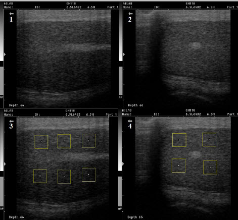
The objective of the present study was to measure pixel intensity mean (Echogenicity) and pixel intensity standard deviation (Heterogeneity) of testicular parenchyma in constantly low or high sperm concentration producing bulls. The average sperm concentration/ml in ejaculate over thirteen month’s period (≥100 ejaculates) was recorded. On the basis of sperm concentration, bulls were grouped into; Low sperm concentration (Group A, n=6: age 4.5 to 6 years) and High sperm concentration (Group B1, n=6: age 4.5 to 6 years and B2, n=3: age 7 to 7.5 years) for this experiment. Digital image analysis of ultrasound scan images was done to measure echogenicity and heterogeneity by using ImageJ software. There was no significant difference in echogenicity values between groups, whereas image heterogeneity values of Group A showed statistically significant (P<0.05) lower in comparison with B1 and B2. However, between Group B1 and B2 there was no difference. No correlation was observed between the echogenicity and heterogeneity values in any of the groups. Heterogeneity of echo structure may indicate the seminiferous tubule diameter, sertoli cell population and fluid density within tubules. In conclusion, lower sperm output was observed in testes that were less heterogenic at the tissue level. The heterogeneity values in bulls for rejection at the time of BSE (Breeding soundness evaluation) should be studied in more detail to have more insight and to incorporate in BSE of Murrah bull.
Downloads
Metrics
References
Arteaga, A.A., A.D. Barth and L.F.C. Brito. 2005. Relationship between semen quality and pixel-intensity of testicular ultrasonograms after scrotal insulation in beef bulls. Theriogenology, 64(2): 408-415. DOI: 10.1016/j.theriogenology.2004.12.008
Barth, A.D., L. Alisio, M. Aviles, A.A. Arteaga, J.R. Campbell and S.H. Hendrick. 2008. Fibrotic lesions in the testis of bulls and relationship to semen quality. Anim. Reprod. Sci., 106(3-4): 274-288. DOI: 10.1016/j.anireprosci.2007.05.002
Bhakat, M., T.K. Mohanty, V.S. Raina, A.K. Gupta and M.H. Khan. 2011. Frozen semen production performance of Murrah buffalo bulls. Buffalo Bull., 30(2): 157-162. Available on: https://ibic.lib.ku.ac.th/e-bulletin/IBBU201102010.pdf
Brito, L.F.C., A.D. Barth, R.E. Wilde and J.P. Kastelic. 2012. Testicular ultrasonogram pixel intensity during sexual development and its relationship with semen quality, sperm production, and quantitative testicular histology in beef bulls. Theriogenology, 78(1): 69-76. DOI: 10.1016/j.theriogenology.2012.01.022
Byrne, C.J., S. Fair, A.M. English, M. Cirot, C. Staub, P. Lonergan and D.A. Kenny. 2018. Plane of nutrition pre and post-six months of age in Holstein-Friesian bulls: I. Effects on performance, body composition, age at puberty and post-pubertal semen production. J. Dairy Sci., 101(4): 3447-3459. DOI: 10.3168/jds.2017-13719
Cardilli, D.J., G.H. Toniollo, A.A. Pastore, J.C. Canola, M.E.Z. Mercadante and J.A. Oliveira. 2010. Padrao ultrassonografico do parenquima, mediastino e tunicas testiculares em bovinos jovens da raca Nelore. Ci. Anim. Bras., 11(4): 899-905. DOI: 10.5216/cab.v11i4.6599
Davies, D.V., T. Mann and L.E. Rowson. 1957. Effect of nutrition on the onset of male sex hormone activity and sperm formation in monozygous bull-calves. In Proceedings of the Royal Society of London Series B-Bio-logical Sciences, UK. p. 332-351.
Eilts, B.E. and R.D. Pechman. 1998. B-mode ultrasound observations of bull testes during breeding soundness examinations. Theriogenology, 30(6): 1169-1175. DOI: 10.1016/0093-691x(88)90292-0
English, A.M, C.J. Byrne, P. Cormican, S.M. Waters, S. Fair and D.A. Kenny. 2018. Effect of early calf-hood nutrition on the transcriptional regulation of the hypothalamic-pituitary testicular axis in holstein-friesian bull calves. Sci. Rep.-UK., 8(1): 16577. DOI: 10.1038/s41598-018-34611-4
Hershkovitz, R., K. Amichay, G.Y. Stein and R. Tepper. 2010. The echogenicity of the normal fetal kidneys during different stages of pregnancy determined objectively. Arch. Gynecol. Obstet., 284(4): 807-811. DOI: 10.1007/s00404-010-1738-0
Ivancic, M. and W. Mai. 2008. Qualitative and quantitative comparison of renal vs. hepatic ultrasonographic intensity in healthy dogs. Veterinary Radiology and Ultrasound, 49(4): 368-373. DOI: 10.1111/j.1740-8261.2008.00383.x
Moxon, R., L. Bright, B. Pritchard, I.M. Bowen, M.B. De Souzac, L.D. Da Silva and G.C. England. 2015. Digital image analysis of testicular and prostatic ultrasonographicechogencity and heterogeneity in dogs and the relation to semen quality. Anim. Reprod. Sci., 160: 112-119. Available on: https://daneshyari.com/article/preview/2072594.pdf
Omer, R., J. Giffin, A. Hahnel and P. Bartlewski. 2012. Relationships of ultrasonographic and magnetic resonance image attributes to the histomorphology of ram testes. Reprod. Biol., 12(4): 355-361. DOI: 10.1016/j.repbio.2012.10.009
Sidibe, M., L.A. Franco, G. Fredriksson, A. Madej and L. Malmgren. 1992. Effects on testosterone, LH, and cortisol concentrations and on testicular ultrasonographic appearance of induced testicular degeneration in bulls. Acta Vet. Scand., 33(3): 191-196. DOI:10.1186/BF03547308
Tomlinson, M., A. Jennings, A. Macrae and I. Truyers. 2017. The value of trans-scrotal ultrasonography at bull breeding soundness evaluation (BBSE): The relationship between testicular parenchymal pixel intensity and semen quality. Theriogenology, 89: 169-177. DOI: 10.1016/j.theriogenology.2016.10.020