Ultrasonographic morphometry of reticulum in cattle and buffaloes suffering from traumatic reticulo-peritonitis

Authors

  • Shanib Mehraj Makhdoomi Department of Veterinary Surgery and Radiology, College of Veterinary Science, Guru Angad Dev Veterinary and Animal Sciences University (GADVASU), Ludhiana, India
  • Vandana Sangwan Department of Veterinary Surgery and Radiology, College of Veterinary Science, Guru Angad Dev Veterinary and Animal Sciences University (GADVASU), Ludhiana, India
  • Ashwani Kumar Department of Veterinary Surgery and Radiology, College of Veterinary Science, Guru Angad Dev Veterinary and Animal Sciences University (GADVASU), Ludhiana, India
  • Jitender Mohindroo Department of Veterinary Surgery and Radiology, College of Veterinary Science, Guru Angad Dev Veterinary and Animal Sciences University (GADVASU), Ludhiana, India
  • Anuradha Gupta Department of Veterinary Anatomy, College of Veterinary Science, Guru Angad Dev Veterinary and Animal Sciences University (GADVASU), Ludhiana, India

Keywords:

buffaloes, Bubalus bubalis, ultrasound, bovine, cow, reticulitis, foreign body

Abstract

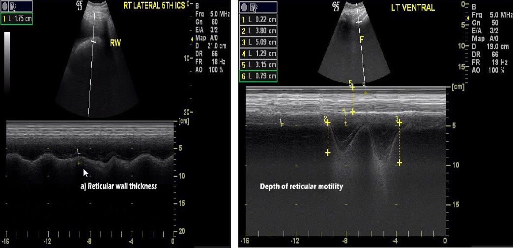
The objectives of the study were to evaluate the ultrasonographic morphometry of the reticulum in healthy non-gravid cattle and buffaloes from right and left parasternal and lateral windows and to evaluate the role of ultrasonography in the prediction of penetrating metallic foreign bodies in the reticular wall in cattle and buffaloes. The study included 22 clinically healthy (11 cross-bred Indian cattle (Bos tarus and Bos indicus) and 11 Indian water buffaloes (Bubalus bubalis)) and 26 traumatic reticulitis affected bovines (21 cattle and 15 buffaloes). Various parameters like, reticular wall thickness, depth of reticulum, pattern of reticular motility and wall, and presence of effusions were recorded to assess the penetrability of metallic sharp foreign body using ultrasonography.
The reticular wall thickness varied from 0.34 to 0.82 cm and 0.37 to 0.68 cm in healthy cattle and buffaloes, respectively. In both the species, the mean highest reticular wall thickness was recorded on the left lateral side. Instead of typical biphasic motility, folding type motility was recorded in 72.73% healthy buffaloes and 27.27% healthy cattle from the left lateral side. The reticular wall pattern was recorded to be smoother in healthy buffaloes from all the windows compared to healthy cattle.
The peri-reticular reaction in diseased bovine was least observed on the left lateral aspect of the reticulum and was maximum evident on the left ventral aspect of reticulum in cattle and the right ventral aspect in buffaloes. The reticular motility was present in maximum number of bovine despite adhesions present on rumenotomy. The cattle showed more adhesions for partially penetrating foreign bodies, compared to buffaloes. Despite non-penetrating foreign bodies, the peri-reticular effusions were seen on the ventral aspect of reticulum from both sides, though in less quantity.
In conclusion, the peri-reticular effusions are maximum seen on the left ventral aspect in cattle and right ventral in buffalo in completely and partially penetrating foreign bodies; however, effusions may also be present in non-penetrating foreign body in cattle.

Downloads

Download data is not yet available.

Metrics

Metrics Loading ...

Author Biographies

Shanib Mehraj Makhdoomi, Department of Veterinary Surgery and Radiology, College of Veterinary Science, Guru Angad Dev Veterinary and Animal Sciences University (GADVASU), Ludhiana, India

MVSc Scholar, Department of Veterinary Surgery and Radiology, College of Veterinary Science, GADVASU, Ludhiana-141004, India

Vandana Sangwan, Department of Veterinary Surgery and Radiology, College of Veterinary Science, Guru Angad Dev Veterinary and Animal Sciences University (GADVASU), Ludhiana, India

Assistant Professor 
Department of Veterinary Surgery and Radiology, College of Veterinary Science, GADVASU, Ludhiana-141004, India

Ashwani Kumar, Department of Veterinary Surgery and Radiology, College of Veterinary Science, Guru Angad Dev Veterinary and Animal Sciences University (GADVASU), Ludhiana, India

Assistant Professor 
Department of Veterinary Surgery and Radiology, College of Veterinary Science, GADVASU, Ludhiana-141004, India

Jitender Mohindroo, Department of Veterinary Surgery and Radiology, College of Veterinary Science, Guru Angad Dev Veterinary and Animal Sciences University (GADVASU), Ludhiana, India

Professor-cum-Head 
Department of Veterinary Surgery and Radiology, College of Veterinary Science, GADVASU, Ludhiana-141004, India

Anuradha Gupta, Department of Veterinary Anatomy, College of Veterinary Science, Guru Angad Dev Veterinary and Animal Sciences University (GADVASU), Ludhiana, India

Professor 
Department of Veterinary Anatomy, College of Veterinary Science, GADVASU, Ludhiana-141004, India

Downloads

Published

2019-10-01

How to Cite

Makhdoomi, S. M., Sangwan, V., Kumar, A., Mohindroo, J., & Gupta, A. (2019). Ultrasonographic morphometry of reticulum in cattle and buffaloes suffering from traumatic reticulo-peritonitis. Buffalo Bulletin, 38(3), 421–436. Retrieved from https://kuojs.lib.ku.ac.th/index.php/BufBu/article/view/562

Issue

Section

Original Article

Most read articles by the same author(s)